cameron ulcer causes
The lesions may cause chronic blood loss resulting in iron deficiency anemia. 3Esophageal gastric and duodenal biopsies were normal and there was no evidence of Helicobacter pylori infection.
2
A lesion associated with large diaphragmatic hernia and chronic.

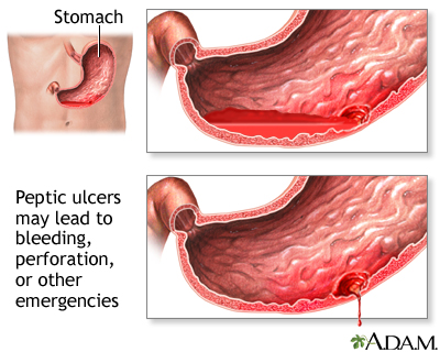
. The lesions may cause chronic blood loss resulting in iron deficiency anemia. More research needs to be performed on surveillance for these patients once a cameron ulcer and lesion has been found to be a cause of overt GI bleeding. Cameron ulcer is a linear gatric ulser on the mucosal folds in patients with a large hiatal hernia.
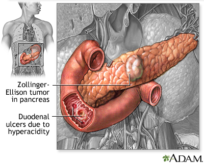
They usually form as a sequelae of an adjacent inflammatory process such as chronic pancreatitis. Previous article Next article Keywords. Zollinger-Ellison syndrome is another condition that can lead to ulcers.
Treatment of anemia with Cameron lesions includes iron supplements and acid suppression by a proton-pump inhibitor PPI. Pylori infection or nonsteroidal anti-inflammatory drugs. Cameron lesions and ulcers are erosions or ulcers on the gastric folds at the level of the diaphragm and can sometimes be seen in patients with large hiatal hernias.
Another rare type of. A Cameron lesion is a linear erosion or ulceration of the mucosal folds lining the stomach where it is constricted by the thoracic diaphragm in persons with large hiatal hernias. This article is part of an expert video encyclopedia.
Less often they cause acute bleeding. It causes mainly iron deficiency anemia due to chronic gastrointestinal bleeding and dyspepsia 2. As our society moves to an ever-increasing national average for BMI bleeding secondary to Cameron lesions should be placed on the differential and an EGD should be carefully performed.
Though typically asymptomatic these may rarely present as acute severe upper gastrointestinal bleed GIB. It is still not clearly understand as to what causes Cameron ulcers however experts believe that it may be associated with irritation caused due to stomach acid mechanical trauma or ischemia due to hampered blood supply to. The lesions are associated with occult bleeding.
9 Other causes could be related to abdominal trauma atherosclerosis other inflammatory conditions or infection embolic events. Surgical hernia repair is sometimes needed. Other complications are mucosal prolapse incarceration volvulus and esophageal shortening 3.
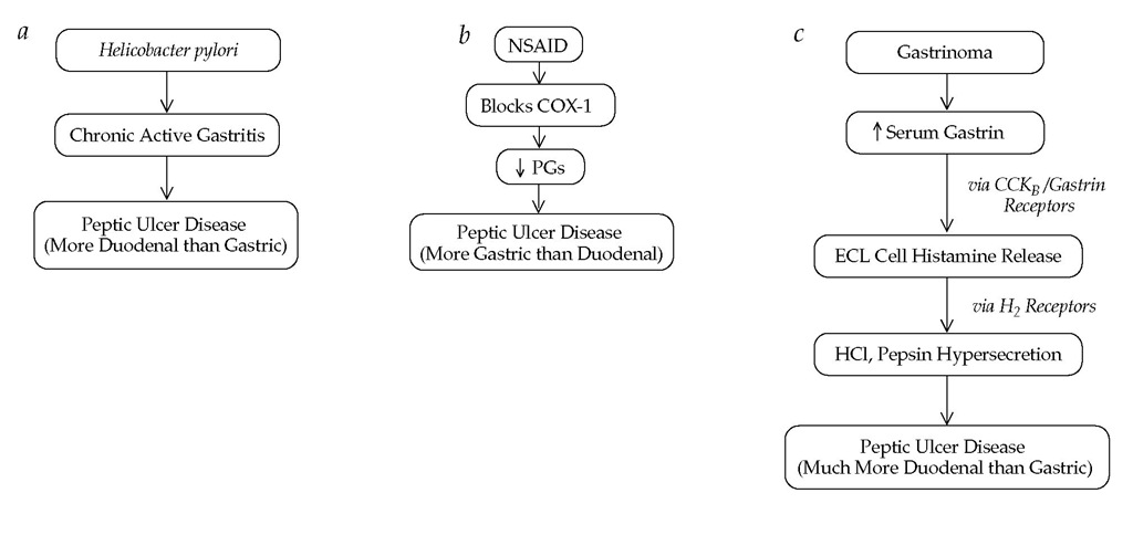
Pylori infection and the use of nonsteroidal anti-inflammatory drugs NSAIDs are the predominant causes of peptic ulcer disease in the United States accounting for 48 and 24 percent of cases. It is found in about 5 of patients with hiatal hernia and sometimes causes acute or chronic upper. Such lesions may be found in upto 50 of endoscopies performed for another indication.
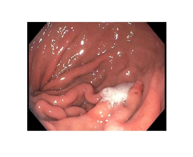
The case authors explain that the pathogenesis of Cameron lesions has not been clearly defined but is thought to be caused by the combined effects of extra- and intra-luminal mechanical and. In fact the initial report of Cameron et al showed a significant improvement of anemia after surgical hernia repair1 References 1. Cameron lesions are linear gastric ulcers or erosions on the mucosal folds at the diaphragmatic impression in patients with a large hiatal hernia.
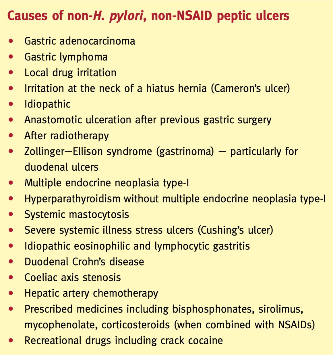
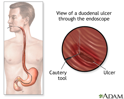
Cameron ulcer could be seen in 5 of patients with hiatal hernia who undergo upper. Causes of peptic ulcers include long-term use of nonsteroidal anti-inflammatory drugs such as aspirin and ibuprofen an infection with the bacteria Helicobacter pylori rare cancerous and noncancerous tumors in the stomach duodenum or pancreas known as Zollinger-Ellison syndrome Sometimes peptic ulcers are caused by both NSAIDs and H. Up to 10 cash back Repeat upper endoscopy demonstrated a large hiatal hernia with fluid retention in the pouch and a 15-cm Cameron ulcer at the diaphragmatic hiatus without stigmata of recent hemorrhage Fig.
It causes gastrinomas or tumors of the acid-producing cells in your stomach which causes more acid. The lesions are associated with occult bleeding and development of chronic iron deficiency anaemia but are often overlooked during routine endoscopy. Bleeding although infrequent occurs from gastric ulceration gastritis or erosions Cameron lesions within the incarcerated hernia pouch.
The Cameron lesions can cause iron deficiency anemia due to acute or chronic bleeding. Ulcers Figures C and D and the patient was referred to undergo surgical therapy fundoplication. Cameron lesions are linear gastric ulcers or erosions on the mucosal folds at the diaphragmatic impression in patients with a large hiatal hernia.
Peptic ulcer disease is a common cause of gastrointestinal bleeding and is usually related to Helicobacter pylori H. The etiology is unknown in about 8 of the cases1 Gastrointestinal bleed is the most common diagnosis among GI-related hospital admissions. The common causes of upper GI bleed in descending order of occurrence are peptic ulcer 38 esophageal or gastric varices 16 esophagitis 13 UGI tract tumor 7 angioectasia 6 Mallory Weiss tear 4 erosions 2 and Dieulafoys lesion 2.
Cameron lesions represent linear gastric erosions and ulcers on the crests of mucosal folds in the distal neck of a hiatal hernia HH. Cameron AJ Higgins JA. Cameron ulcer kamĕr-ŏn -rŏn An ulcer or a linear erosion found in a hiatal hernia.
Respiratory complications can result from mechanical Evaluation of suspected small bowel bleeding formerly obscure gastrointestinal bleedingnot reached. Patients with a large hiatal hernia are at risk for a Cameron ulcer which has a different physiology and treatment options. A Cameron lesion is a linear erosion or ulceration of the mucosal folds lining the stomach where it is constricted by the thoracic diaphragm in persons with large hiatal hernias.
The most common causes of upper GI bleed include peptic ulcer disease gastroesophageal varices esophagitis angioectasia and vascular lesions. Cameron erosion and ulcers represent the mild and sever form of the same disease spectrum respectively 2.

Cameron Ulcer Causing Severe Anemia In A Patient With Diaphragmatic Hernia Abstract Europe Pmc

Helicobacter Pylori Infection And Peptic Ulcers Medicine
Cameron Lesions And Disorders Of The Cardia Human Anatomy Nuclear Imaging Digestive System

Pdf Evaluation Of Occult Gastrointestinal Bleeding Semantic Scholar
Peptic Ulcers Smartengage

Gastric Ulcers Iv The Gastrointestinalatlas Gastrointestinalatlas Com
Causes Of Peptic Ulcer Disease H Pylori Nsaids Grepmed
In Depth Reports Peptic Ulcers
In Depth Reports Peptic Ulcers

Comparision Of Patients With Ulcer Bleeding And Variceal Bleeding Download Scientific Diagram
Cameron Lesions Article

Peptic Ulcer Chapter Closed Semantic Scholar

Peptic Ulcer Disease Gi Pocket Medical Knowledge Facebook
Peptic Ulcer Diseases Part 1

American Gastroenterological Association Aga In A New Clinical Guideline Aga Recommends Bidirectional Endoscopy For Most Patients With Iron Deficiency Anemia Early Gastrointestinal Evaluation Can Lead To The Identification And Treatment Of

Hiatal Hernia Vs Ulcer What You Need To Know The Surgery Group
In Depth Reports Peptic Ulcers

Cirbosque Some4surgery On Twitter Pictorial Explanation Of The Cameron Ulcers In A Paraesophageal Hernia By Dr Frank Netter Some4surgery Ascolcx2020 Medtwitter Juliomayol Swexner Pferrada1 Pipecabrerav Almagoch Drthawaba Neilflochmd

What Are Cameron Lesions Causes Symptoms Treatment Natural Remedies


Comments
Post a Comment